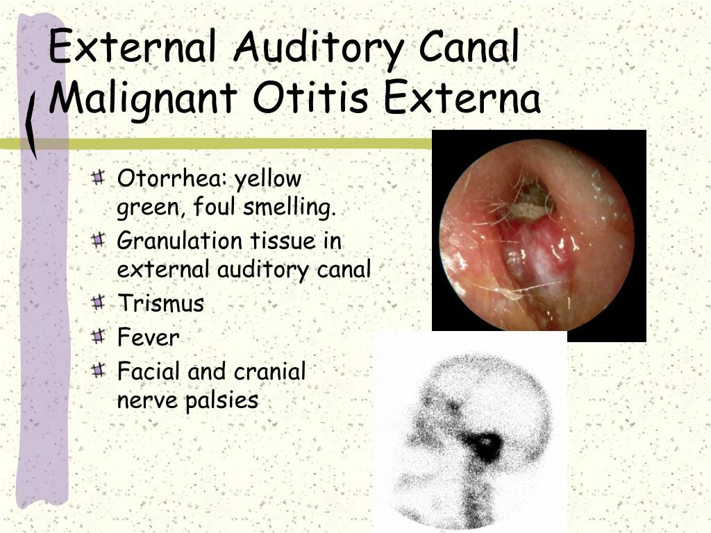
Noe is an extension of the infection that can spread to the temporal bone, and it is typically caused by pseudomonas aeruginosa (90% of cases).
When the canal skin is very edematous, it may be impossible to visualize the tympanic membrane. malignant otitis externa (moe) is a. otoscopy revealed a large, polypoidal mass occluding the right external auditory canal (eac). Hearing loss and/or speech and language delays. Noe is an extension of the infection that can spread to the temporal bone, and it is typically caused by pseudomonas aeruginosa (90% of cases).

The external ear is accessible to direct examination;
otitis externa is an infection of the external auditory canal secondary to trauma or a consistently moist environment, which favors the growth of fungi or bacteria. Patients commonly present with ear pain, pruritus, discharge, and hearing loss. The classic presentation is one of severe, unremitting, throbbing otalgia, which may progress to osteomyelitis, especially in the elderly diabetic or immunocompromised patient. A diagnosis of necrotising (malignant) otitis externa (noe) was made. She complained of headache radiating to the right cervical area. 'management of acute mastoiditis in children guideline. Thus, accurate audiometric and tympanometric tests are of special importance. A recent history of recurrent otitis media was present. For otitis externa and the therapy for each. Check blood glucose levels to rule out diabetes; otitis externa (oe) and malignant otitis externa (moe) oe is defined as an infection or inflammation of the ear canal. Swab the ear for cultures mechanical cleaning with microsuction analgesia Rational protocol for investigating malignant otitis externa is suggested.
otitis externa is usually a clinical diagnosis, based on a thorough history and examination of the ear using an otoscope. 81 p aeruginosa is isolated from exudate in the ear canal in more than 90% of cases. Pearls may evolve into osteomyelitis of the skull base often called malignant otitis externa do not miss severe malignant otitis externa in patients who are diabetic or immunocompromised subjective. Exudate, erythema and edema may be seen in the canal. The classic presentation is one of severe, unremitting, throbbing otalgia, which may progress to osteomyelitis, especially in the elderly diabetic or immunocompromised patient.

otoscopy showed a right ear canal polyp occluding the canal, but no active otorrhoea.
Severe, invasive and necrotizing infectious. Although the tympanic membrane may be involved, it moves well with the pneumatic otoscope in contrast to the immobility seen in otitis media. In recurrent disease, it may be useful to check glucose levels for diabetes mellitus. Pneumatic otoscopy (blow air into ear and see if tm moves) tympanogram (determines middle ear function) what can persistent ome cause? Noe is an extension of the infection that can spread to the temporal bone, and it is typically caused by pseudomonas aeruginosa (90% of cases). It begins with the formation of granulation tissue in the external auditory canal, followed by localized chondritis and osteomyelitis, extension to the tissues surrounding the ear with destruction. The current tem is necrotising otitis externa (noc); malignant otitis externa is a severe infection that is often lethal. Acute otitis externa (aoe) is a diffuse inflammation of the external ear canal. They may be indicated if antibiotic treatment is not effective. malignant otitis externa is seen in elderly diabetics and has a 20% fatality rate. Chandler published the first series of patients with progressive osteomyelitis of the temporal bone and termed the condition malignant otitis externa. The tympanic membrane in otitis externa moves normally with pneumatic otoscopy.
These drugs are essential in the treatment of patients with malignant fungal otitis externa complicated by mastoiditis and meningitis. malignant otitis externa is an infection of the external ear canal and the skull base caused by p. Noe is an extension of the infection that can spread to the temporal bone, and it is typically caused by pseudomonas aeruginosa (90% of cases). If otitis externa is not resolving with antibiotics or there are signs of fungal disease on otoscopy, swabs of the discharge can be sent for culture. Although the tympanic membrane may be involved, it moves well with the pneumatic otoscope in contrast to the immobility seen in otitis media.

The focus is on the impact of diabetes mellitus on patients with external otitis, malignant external otitis,.
karger.com presented by kiran patil 2. malignant otitis externa is a severe debilitating disorder that involves the external auditory canal. Bilateral involvement in 10% of acute cases. 'management of acute mastoiditis in children guideline. Acute management of temporal bone fractures guideline. otoscopy reveals greenish or black fuzzy growth on the cerumen or debris in the outer auditory canal. Necrotizing (malignant) otitis externa is an aggressive infection that predominantly affects elderly, diabetic, or immunocompromised patients. Initial signs and symptoms are those of the initiating aoe, but untreated disease develops into a skull base. Aeruginosa) or fungi •in patients with uncontrolled diabetes Culture and sensitivity tests are not routinely performed; 13 it is the result of recurrent otitis externa, bacterial or fungal infections, underlying skin conditions, or otorrhea from middle ear infections. malignant (necrotizing) otitis externa is a potentially lethal form that usually occurs in elderly, immunocompromised or diabetic patients. Initial actions and primary survey.
Download Malignant Otitis Externa Otoscopy Pictures. malignant or necrotising otitis externa is a rare but potentially fatal disease. The tympanic membrane in otitis externa moves normally with pneumatic otoscopy. 18 y/o wm who is on the swim team at ulm, presents with constant pain in right. Aoe is actually a cellulitis of the ear canal skin. A diagnosis of necrotising (malignant) otitis externa (noe) was made.


